
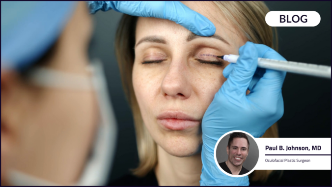
Upper Blepharoplasty: Functional Gain, Tissue Preservation and the Discipline of Millimeters
When patients say, “I just want my eyes to look less tired,” upper blepharoplasty is often perceived as cosmetic. In practice, functional concerns frequently drive consultation — and objective evidence supports measurable improvement in visual field and quality of life following surgery.
More than 120,000 blepharoplasties were performed in the United States in 2024 alone. For many patients, redundant upper eyelid skin contributes to superior visual field obstruction and compensatory brow elevation.
A 2011 American Academy of Ophthalmology (AAO) assessment reported significant improvement in vision, peripheral vision and quality-of-life activities following functional upper blepharoplasty and ptosis repair.
To understand how those outcomes are achieved surgically, we spoke with Dr. Paul Johnson, a board-certified oculofacial plastic surgeon, about how he evaluates obstruction, plans excision and approaches tissue balance.
Objective Evaluation: Defining the Source of Obstruction
Dr. Johnson begins with measurement:
- Margin reflex distance (MRD)
- Levator function
- Brow position
- Dermatochalasis and fat prolapse
These measurements guide surgical planning and insurance documentation and help distinguish dermatochalasis from brow descent or true ptosis.
He notes that patients frequently describe the need to raise their brows in order to see — a compensatory behavior that often correlates with measurable superior field limitation.
Clinical Context: Blepharoplasty as a Millimeter Procedure
Upper eyelid surgery is frequently described as a millimeter-level operation. Small variations in crease height, skin preservation or muscle handling can influence eyelid closure mechanics, corneal coverage and aesthetic symmetry.
Functional outcome studies demonstrate measurable expansion of superior visual field after appropriate tissue excision. At the same time, ocular surface research indicates that surgical technique and extent of tissue removal may influence tear film stability in certain patients, particularly in the early postoperative period.
Longitudinal data suggest no significant long-term worsening of dry eye symptoms for most patients following upper blepharoplasty. However, early changes in tear breakup time or surface parameters have been observed in controlled settings.
Taken together, the literature reinforces a central surgical principle: functional gain must be balanced against preservation of blink dynamics and surface protection.
Surgical Execution: Planning for Balance
In his own practice, Dr. Johnson describes a measured approach. For most patients, he creates a lid crease approximately 7 mm above the lid margin and aims to preserve roughly 13 mm of skin — a balance he finds optimizes both functional improvement and aesthetic harmony.
He performs a final intraoperative re-draping check before closure to confirm adequate preservation and avoid over-resection. In millimeter-level surgery, clean incisions and controlled tissue handling support reproducible symmetry.
From a surgical standpoint, this type of restraint reflects an understanding that eyelid tissue is not redundant by default — it contributes to closure force distribution, tear film spread and corneal protection.
Precision in incision placement and tissue handling therefore sets the foundation for both exposure and protection.
Ocular Surface Risk: Patient Selection and Monitoring
Dry eye symptoms are a common preoperative concern in eyelid surgery.
Evidence indicates stable long-term dry eye outcomes in most patients. At the same time, studies examining technique variations demonstrate transient changes in ocular surface parameters depending on tissue handling, underscoring the importance of baseline assessment.
Dr. Johnson notes that patients with preexisting surface symptoms warrant careful evaluation and follow-up.
Anatomical Variation and Ethnic Considerations
Crease height and tissue configuration vary by anatomy and patient preference.
Dr. Johnson notes that many Asian patients prefer outcomes that preserve their natural ethnic eyelid characteristics rather than adopting a higher crease configuration. Individualized planning remains central to surgical execution.
Communication as Part of the Outcome
Functional improvement does not automatically translate to patient satisfaction.
Dr. Johnson emphasizes unhurried consultation, explanation of measurements, and realistic expectation-setting. For patients undergoing surgery for visual obstruction, he often describes the result as “raising a window shade” — expanding the visual field and allowing more light and clarity.
Minor refinements, when necessary, are typically reassessed after full healing.
Precision as a Clinical Discipline
Upper blepharoplasty sits at the intersection of anatomy, function, and surgical judgment.
Evidence supports:
- Visual field expansion and improved quality of life
- Stable long-term dry eye outcomes in most patients
- Technique-sensitive, often transient changes in tear film parameters
For surgeons, the technical challenge is not simply removing tissue — it is removing enough to relieve obstruction while preserving enough to protect the ocular surface.
In that sense, upper blepharoplasty exemplifies a broader surgical discipline: millimeters matter.